Electron Microscopy Core
The Electron Microscopy Core is part of the Biology Advanced Imaging Core.
Overview
There are two main instruments, a 125keV instrument for routine electron microscopy and a 200keV instrument for 3D tomography and for cryo-EM of protein structure.
See the Introduction to Electron Microscopy Tutorial
Hitachi 125keV H-7100 Transmission Electron Microscope
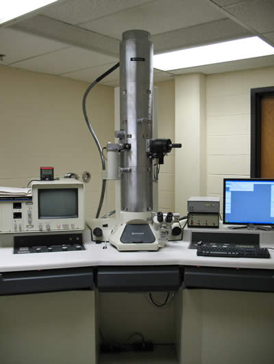
This instrument is excellent for standard electron microscopy needs to image sections between 33nm to 70nm thick. with a Gatan Orius SC1000 slow scan 4kX2.6k CCD camera for acquisition of high resolution digital images.
Features:
- Tungsten filament or LaB6
- Point resolution: 0.3 nm
- Information limit: 0.14 nm
- Magnification range: 1k-500kx
- GATAN Orius SC1000 digital camera: 4008 x 2672 pixels high resolution camera
- GATAN digital micrograph for computer assisted mosaic imaging
- Rotation and tilt specimen holders
FEI 200keV Tecnai G2 F20 TWIN Transmission Electron Microscope
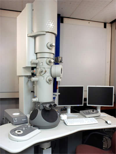
This instrument is a high-end instrument capable of operating at voltages between 40keV and 200keV to image samples of varying thickness. Tissue sections up to 200nm thick can be imaged. 3D images can be computed by tomography; the stage can rotate ±70°. This microscope can acquire high resolution cryo-electron microscopy images of protein complexes. Fitted with an Eagle 2kX2k CCD camera for acquisition of digital images.
The installation of the FEI Tecnai G2 F20 was completed May 2011.
Features:
- ZrO2/W (100) Schottky Field emitter (FEG)
- TWIN objective lens with computerized 5-axes eucentric goniometer
- Line resolution at zero tilt: 0.23 nm
- Line resolution at 45º tilt: 0.34nm
- FEI Eagle HR CCD camera: 2k x 2k CCD camera
- High-resolution and high dose quantum efficiency
- Bottom mounted (cannot mount a post-column energy filter)
- CCD pixel size: 30 x 30 µm^2
- Readout speed: 2.5 Mpixel/sec
- Single and double tilt specimen holders (α= +/- 70º)
- Gatan 70 degree Cryo-Transfer holder and Cryo-Test with Cryo Box
- Xplore3D for automated tomographic tilt series acquisition
- Inspect 3D on a support PC for tomographic reconstructions
- Amira visualization software
High-pressure freezing
See the High-Pressure Freezing Tutorial.
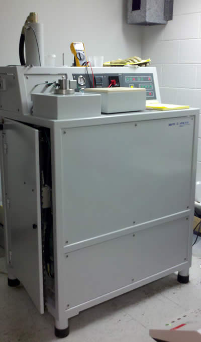
Leica EMPact2 High Pressure Freezing Machine. (RTS not included)
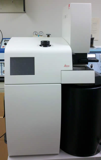
The EMPact 2 was originally designed for time-resolved electron microscopy by allowing users to transfer specimens from a light microscope to the freezer in less than 2 seconds. This device is modified to allow light stimulation of the sample at time points ≥20ms before freezing. Coupling light-activatable molecules or optogenetic tools with high-pressure freezing, morphological changes at an ultrastructural level can be studied.
Features:
- Home-built programmable light stimulation device with 5 ms temporal resolution
- Automated light application and freezing
- 2000 bar pressure applied to samples in 5ms
- -25,000ºC/s cooling rate
- Temperaure/pressure curve can be stored and exported
- Sample as thick as 200µm can be frozen
- Various specimen carriers available
- Low liquid nitrogen consumption
- Low noise
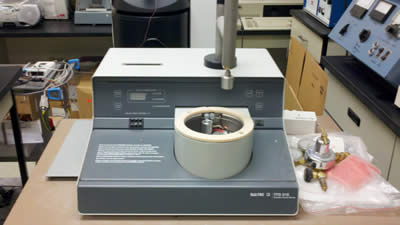
Bal-Tec HPM 010 High Pressure Freezing Machine
This device produces high quality freezing of tissue samples with up to 300µm thickness. HPM 010 is the first commercially available high-pressure freezer, yet it is still the most reliable and robust freezing device.
Features:
- Working pressure range: 2300 – 2600 bar
- Liquid nitrogen and pressures are applied to both sides of the specimen, allowing uniform freezing of specimens
- Specimens as thick as 300 µm can be frozen
- Cooling time from 0ºC to -50ºC: 10 ms
Plunge freezing
A simple and inexpensive way to freeze tissues thinner than 1 µm is plunge freezing. In this technique, specimens are immsersed into liquid propane. Liquid propane has a cooling rate of ~12,000ºC/s. At this freezing rate, the surface of specimens can be frozen without ice crystals. Thus, it is suitable for cell cultures.
BalTec TFD 010 Transfer Freeze Device
This device freezes the surface of tissues up to ~500 nm. It is often used to prepare the samples for freeze-fracture.
Features:
- Inexpensive and simple to use
Freeze-substitution
To examine frozen tissues at room temperature, vitrified ice are exchanged with organic solvent such as acetone at low temperature (-90º). Specimens can then be brought up to room temperature without ice crystals. At the same time, tissues are fixed, often, with osmium tetroxide, which cross-links lipids and provides contrast in tissues under an electron microscope.
Leica AFS automatic freeze substitution machine
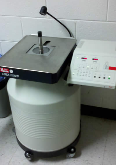
This device is capable of holding temperatures ranging from -140ºC to 60ºC with the use of liquid nitrogen. Speed of temperature change can be controlled with the minimum speed of 1ºC/hour.
Features:
- Temperaure control from -140ºC to 60ºC
- 10 programs can be stored
- UV lamp for plastic polymerization
Fixation, microtomy and staining
Pelco 3450 Laboratory Microwave Processor
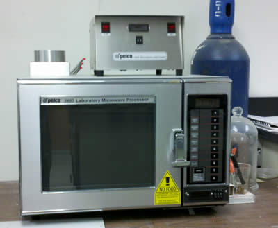
The power of microwave in your household is controlled by changing the intervals between microwave application and rest. This processor allows continuous power outputs ranging from 100 to 750 W. The microwave processing at low power promotes diffusion of chemicals without damaging tissues, and thereby speeds up processes such as immunolabeling, fixation, and infiltration that typically require tremendous time.
Features:
- The power output can be varied from 100 to 750 W
- Continuos power can be applied to specimens
- Program can be stored
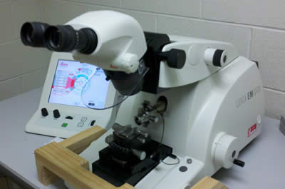
Leica EM UC6 ultramicrotome
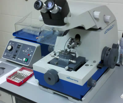
Reichert Ultracut E ultramicrotome
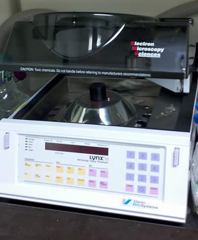
EMS Lynx Microscopy Tissue Processor