
Building a Scanning PALM Microscope
The Theory
The diffraction limit of light was once considered unbreakable; it is a result of the wave-like nature of light itself. Within the past 5 years, this barrier has been broken, opening new possibilities for visualizing molecules at a nanometer scale. This breakthrough has let loose a revolution in optical microscopy -- super-resolution techniques can circumvent the diffraction limit of light. Super-resolution optical imaging includes point-spread function engineering (STED [1]), and computational localization techniques (PALM [2], FPALM [3], BP-FPALM [4], and DH-PSF [5]).
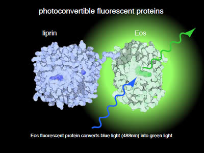
Figure 1: For PALM microscopy proteins can be tagged with a photoconvertible fluorescent protein. Initially this protein emits green light when exposed to blue light.
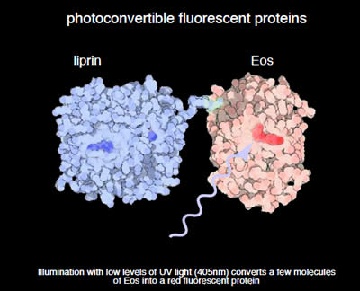
Figure 2: Exposure to a low level of UV light will convert just a few molecules to a red emitting fluorescent protein. The converted molecules should be spaced by greater than the diffraction limit from each other.
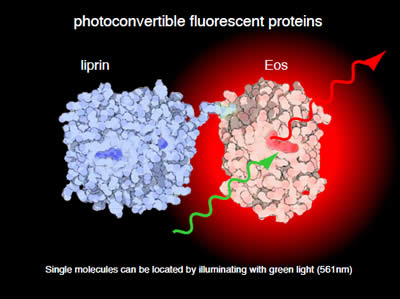
Figure 3: These molecules can now be stimulated with green light and emit red light. The camera records a diffraction-limited spot, but the computer can calculate the center of the spot. Eventually the fluorescent protein is bleached and is effectively erased from the field. The process is repeated until all of the molecules are localized.